arivis Scientific Image Analysis

Image Analysis Solutions

Our software ecosystem enables you to automate your image analysis tasks - from routine applications to challenging deep-dive analyses. Learn more about some of our solution examples below.

nuclei_and_micronuclei

Nuclei and Micronuclei Analysis

Analyze the volume and number of nuclei and micronuclei in whole embryos

Read more

cell_tracking

Cell Tracking

Segment and track individual cells based on phase contrast imaging to analyze cell motility

Read more

DNA_foci_assay

DNA Foci Assay

Hierarchical parent/child analysis of foci within nuclei  

Read more

high-content-genotoxicity-1

High-content Genotoxicity

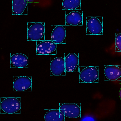
Quantify the level of DNA damage in DAPI stained cells by measurement of signal intensities of foci in 2 channels                                                                                                                               

Read more

dendritic_spine_analysis

Dendritic Spine Analysis

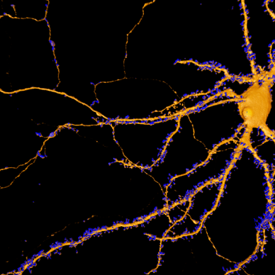
This analysis involves examining dendritic spines and neuronal projections to understand neural circuits                                                                             

Read more

phenotypic_screening

Phenotypic Screening

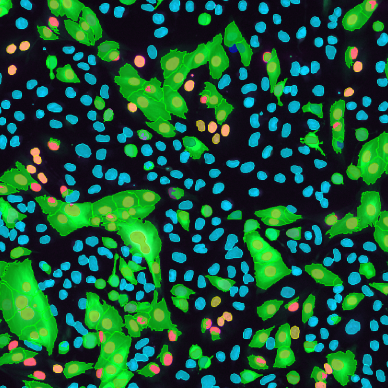
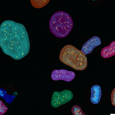
Phenotypic characterization of cells based on cell morphology and intensities of multiple fluorescent markers in the nucleus, cytoplasm or membrane

Read more

organoid_volume_quantification

Organoid Volume Quantification

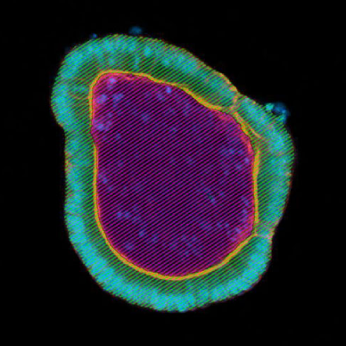
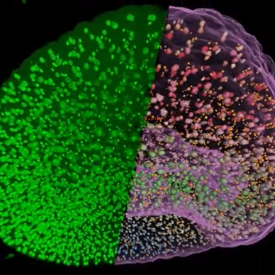
Analyze the volume of organoids and their lumen to quantify growth and differentiation                                                

Read more

cell_level_organoid_analysis

Cell-level Organoid Analysis

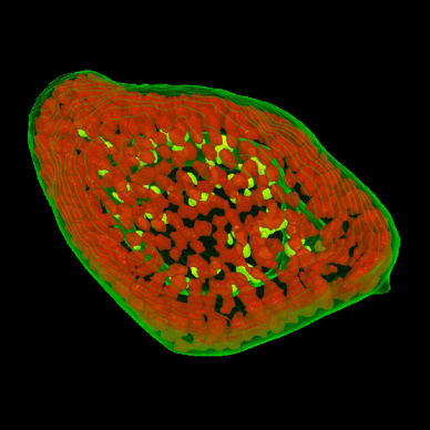
Segment single cells within organoids in 3D to quantify cell numbers and marker expression to analyze organoid growth and differentiation

Read more

nuclei_counting

Nuclei Counting

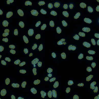
Based on a fluorescent nuclear marker such as DAPI, this solution counts the number of nuclei         

Read more

cell_counting

Cell Counting

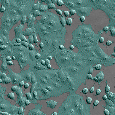
This solution counts the number of cells in phase contrast microscopy images

Read more

cell_confluency

Cell Confluency

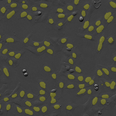
This solution quantifies the area covered by cells in phase contrast images

Read more

talk_to_an_expert

Talk to an Expert

These solution examples got you thinking about your own image analysis challenges?

Contact us

Yes - I want to Talk to an Expert

I want to learn more about ZEISS arivis solutions