arivis Scientific Image Analysis

Nuclei and Micronuclei Analysis in Embryos

Contact us


Nuclei and Micronuclei Analysis in Embryos


Color B/W

Before and After Analysis

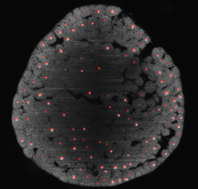
Interact with the image to see before and after.

Adjust the slider to view the 'before and after' comparison.

Solution Description

Manual segmentation of micronuclei and nuclei in 3D is an extremely tedious task even for just one entire embryo. Automated segmentation and analysis of these types of nuclei opens up the possibility to analyze genotoxic stress and the effect of therapeutic drugs. 


64a6eeb47ff2bd5582c07a7c_three dimensional view3

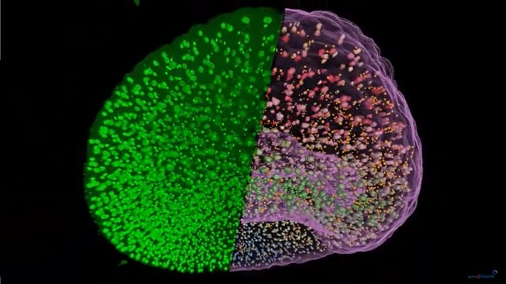
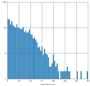
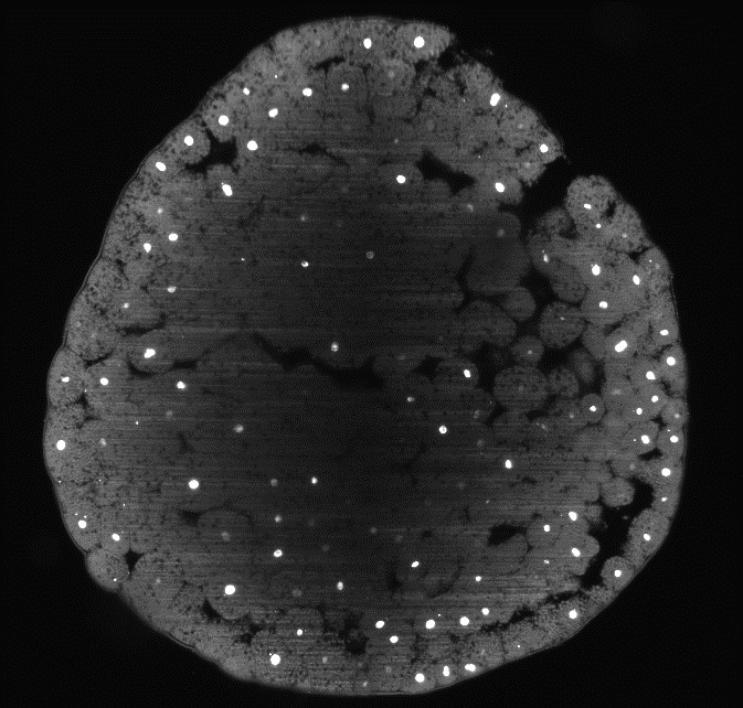
This solution can be used to quantify the number and volume of nuclei and micronuclei separately. First, a deep learning model is used to segment the micronuclei and nuclei in entire z-stacks acquired of whole embryos. Then, 3D analysis is done to quantify features such as the number and volume of nuclei and micronuclei. The results are visualized in graphs such as the histograms shown below.

64a6ef415ed268170217cfff_volume distribution of nuclei

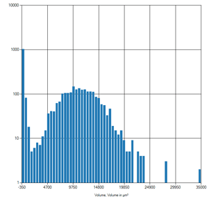
Histogram of nuclei volume.

64a6efab0347dde0e074e9a4_volume distribution of micronuclei

Histogram of micronuclei volume.
Keywords:
deep learning, arivis Cloud, arivis Cloud, arivis Pro, arivis Vision4D, embryo, nuclei, micronuclei, 3D