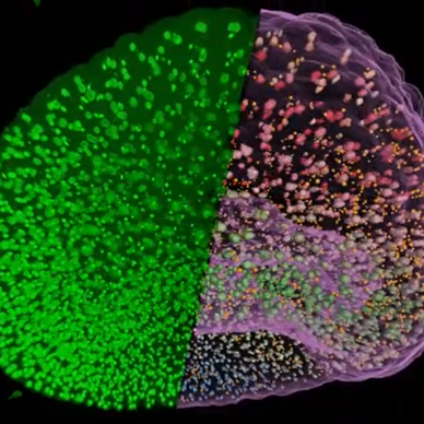
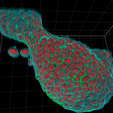
Analyze the volume and number of nuclei and micronuclei in whole embryos

Segment and track individual cells based on phase contrast imaging to analyze cell motility

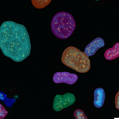
Hierarchical parent/child analysis of foci within nuclei

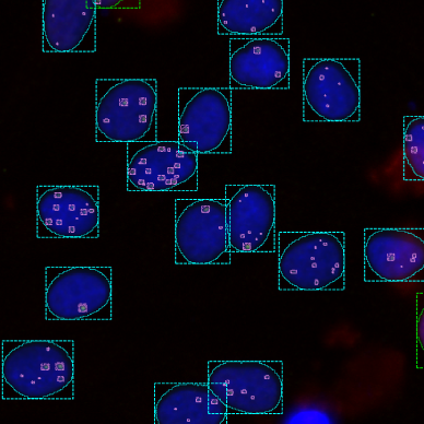
Quantify the level of DNA damage in DAPI stained cells by measurement of signal intensities of foci in 2 channels

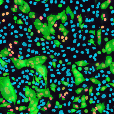
Phenotypic characterization of cells based on cell morphology and intensities of multiple fluorescent markers in the nucleus, cytoplasm or membrane

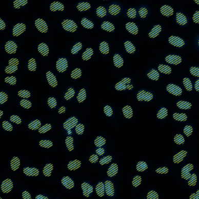
Based on a fluorescent nuclear marker such as DAPI, this solution counts the number of nuclei

Learn about automated, scalable AI Image Analysis for Volume Electron Microscopy to analyse mitochondria in the nucleus and the nuclear membrane and pores.

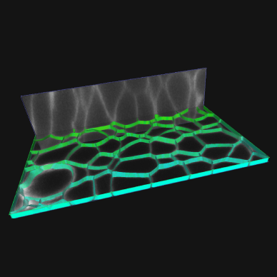
Finding cell boundaries based on membrane-localized contrast is made many times easier by new algorithms that enhance membranes in 3D and an operator designed to segment cells in 3D.

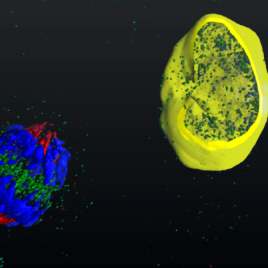
The Colocalization operator is ideal for finding relationships between objects, in a one to any relationship.

The segment tracker operator used for connecting moving objects in time, that is finding relationship of objects during a time course.

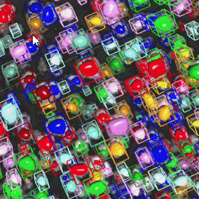
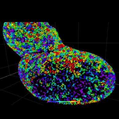
The Blob Finder analysis operator is ideal for segmenting cells or any other type of rounded cell organelles in a noisy image in an easy-to-use 3-step process.

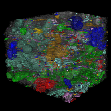
Identifying objects in images acquired by electron microscopy (EM) can be challenging. Since contrast and intensity distributuion in EM images is generally low, simple segmentation algorithms which are based on intensity thresholds or contrast detection often fail with such datasets.

Tracking cells or subcellular particles in microscopic data can be challenging. The Tracking Module allows analysis of the movement of small or large objects over time in both 2D or 3D multichannel image sets of any size.

High-Content Analysis (HCA) has played a significant role in infectious disease research and drug discovery to date and can be a strong tool in the age of COVID-19 and beyond.

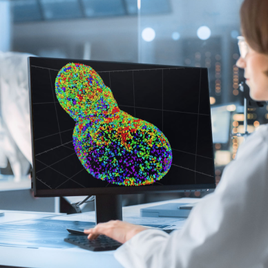
These solution examples got you thinking about your own image analysis challenges?

