Finding cell boundaries based on membrane-localized contrast is made many times easier by new algorithms that enhance membranes in 3D and an operator designed to segment cells in 3D.
This new workflow allows use of any image filters upstream of the Membrane-based Segmentation, which enables optimization of results and/or application to challenging images.
Users can select between Membrane Enhancement and Objectness Measure (sheets) filters depending on data quality and time constraints. Membrane Enhancement gives the highest quality but takes longer while Objectness Measure computes faster. All the other filters in ZEISS arivis Pro (formerly Vision4D) - Morphology, Denoising, etc. - are at the users disposal for fine tuning.
The Membrane-based Segmentation is adjusted interactively to increase cell segmentation yield and “over splitting” is easily proof-edited with the Merge tool.
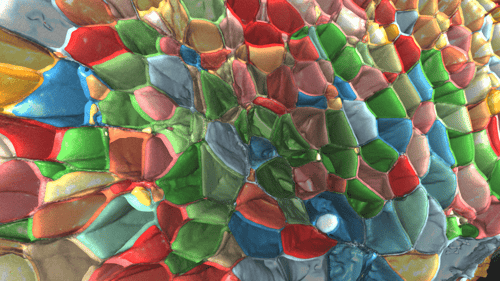
Image courtesy: These data were kindly provided by Tsung-Li Liu, Srigokul Upadlyayula, Tom Kirchhausen, and Eric Betzig and appear in their excellent paper: “Observing the cell in its native state: Imaging subcellular dynamics in multicellular organisms”
Find out how you can enhance your membrane stains with ZEISS arivis Pro (formerly Vision4D).
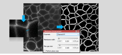
Measure the width of membranes and optimize the voxel filters in your pipeline.
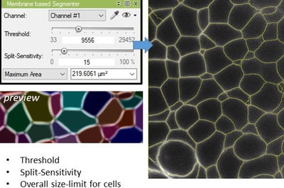
Adjust the Threshold to define the borders of cells and tune the sensitivity to increase or decrease 3D splitting of cells.
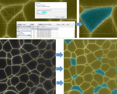
Fix over-splitting via the Merge Objects tool.
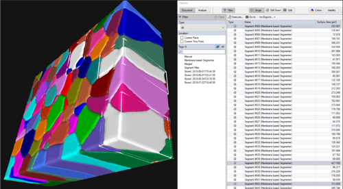
The Objects database in ZEISS arivis Pro (formerly Vision4D) calculates measurements based on the segmented objects. You can highlight individual or groups of cells interactively in the Objects table, or in the 2D / 4D Viewers.
Measure and sort by myriad features such as volume, sphericity, mean intensity and more.
Export the relevant measurements to Excel or CSV for any other software for additional analysis.
Learn more about ZEISS arivis Pro ->