arivis Scientific Image Analysis

DNA Foci Assay

Contact us


DNA Foci Assay


Color B/W

Before and After Analysis

Interact with the image to see before and after.

Adjust the slider to view the 'before and after' comparison.

Solution Description

Analysis of foci within nuclei is important for assays such as a DNA damage assays, fluorescence in situ hybridization (FISH) assays, cell division assays, or any other assay that involves structures visible as foci (also referred to as punctae or spots) within the nuclei.


63e37c9e544eeac28651df56_3D original-1

Figure 1: 3D representation of original image. Nuclei in blue and foci in red.

63e37cd279be5130e2cf6efd_3D Analysis-1

Figure 2: 3D represetation of segmented foci (yellow) inside nuclei (random colors).

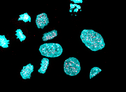
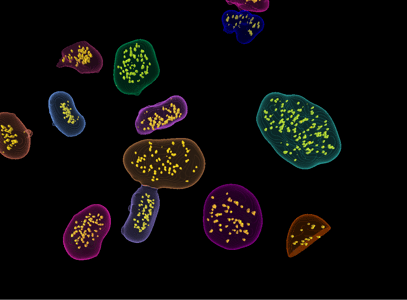
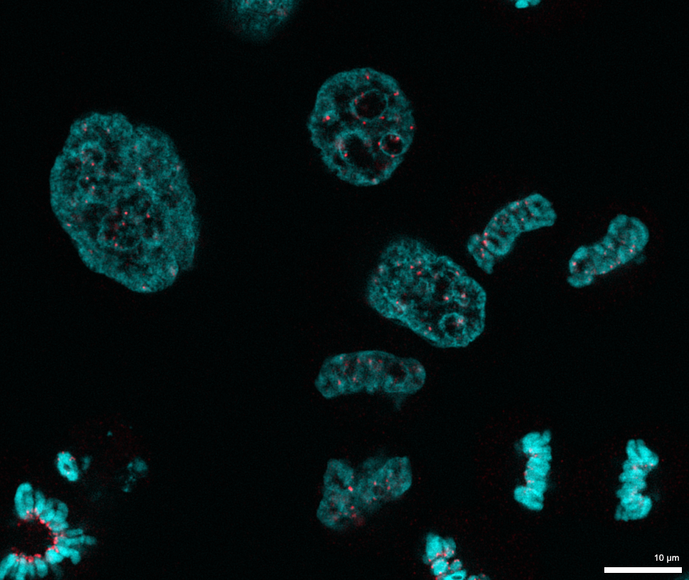
Calculate the number, volume, intensity, density of such foci contained within nuclei. The nuclei and foci are each segmented separately in 3D. A compartments operation then allows for analyzing parent child relationships to quantify foci contained within each nucleus. The user can specify if the foci must be within, overlap or within a certain distance of the nuclear boundary.
 
Keywords:
Parent/child relationships, compartments, foci, punctae, spots, DNA damage, DNA repair, FISH, arivis Vision4D
 
Image Source:
Imaging was performed by Uros Krzic, ZEISS Microscopy.