arivis Scientific Image Analysis

Importing Labelled Images From Open Source

Contact us

The most flexible way to import & export imaging data & results

With arivis you can now integrate all tasks of image visualization and analysis of multidimensional microscopy imaging data of any size in a single software package.

The Open Source community offers many special functionalities and regularly adds new ones to the research community. On the other hand many users prefer a commercial, robust and fully supported image analysis platform with a unified UI that covers all aspects of image processing, analysis and visualization of results via desktop workstation, cloud computing or Virtual Reality. Therefore an easy transition between open source/third party packages and our arivis Scientific Imaging Platform is essential.

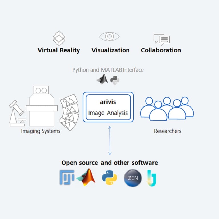
With the ZEISS arivis Pro Exchange Objects with Open Source module we offer an integrated solution to imaging facilities and PIs to connect with open source workflows as well as with any other software which is capable of exporting/importing labelled images. Easily interface with your specific workflows whenever you are using segmentation functions with open source packages such as Fiji, ImageJ, Ilastik, Python or with commercial solutions such as MATLAB or ZEN/APEER software by ZEISS Microscopy.

No programming knowledge needed - ZEISS arivis Pro adapts to your requirements and workflows!

Importing images from open source

Contact us

Importing Labelled Images Workflow

arivis-exchange-objects-workflow-imaging-data-vision4d

1
1. Acquire Acquire your raw microscopy data
2
2. Create

Perform segmentation with your preferred scientific open source or commercial package

3
3. Export Export segments as labeled images
4
4. Analyze Convert labeled images into 3D objects to create 3D movies and enable further analysis 
WEBINAR

Use Machine Learning-based segmentation in whole mouse brain light sheet data

Constanze Depp from the Max Planck Institute of Experimental Medicine is presenting a whole brain quantification of amyloid plaque load using Machine Learning in ZEISS arivis Pro (formerly Vision4D).
Talk to an Expert
Video Tutorials

Import Objects from Open Source

Import from open source or other software into ZEISS arivis Pro (formerly Vision4D) and create impressive movies

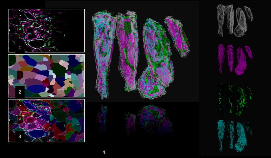
Import resulting objects e.g. from Fiji or any other software in a single step into ZEISS arivis Pro (formerly Vision4D) and visualize the segmentation in 3D to create impressive movies with our arivis storyboard. This is a combined workflow using Ilastik autocontext, 3D segmentation using ImageJ-Matlab. 

Image courtesy of Dr. Carlo Beretta, Livia Asan, Dr. Johannes Knabbe and Prof. Dr. Thomas Kuner, Department of Functional Neuroanatomy, Heidelberg University

Explore & edit resulting objects from open source and ZEISS arivis Pro (formerly Vision4D) in arivis Pro VR (formerly VisionVR)

Import resulting objects e.g. from Fiji or any other software in a single step into ZEISS arivis Pro (formerly Vision4D), then explore and edit the results from open source in immersive Virtual Reality with arivis Pro VR toolkit (formerly VisionVR). This is a combined workflow using Ilastik auto context, 3D segmentation using ImageJ-Matlab.

Image courtesy of Dr. Carlo Beretta, Livia Asan, Dr. Johannes Knabbe and Prof. Dr. Thomas Kuner, Department of Functional Neuroanatomy, Heidelberg University

Full Integration in ZEISS arivis Pro

The one-step import and export of objects allows to add more functionality from Open Source or other microscopy software packages to the proven 3D visualization and analysis functionality of your arivis Scientific Imaging Platform, making it the most flexible solution on the market. No specialized coding/programming skills required!

Your benefits:

  • An easy workflow integrating functionality offered by other software packages
  • Create high quality movie renderings for your publications 
  • Visualize results in synchronized 2D/3D split views
  • Visualize results with feature-based color coding or interactive gating in scatter plots or time line graphs
  • Explore and further edit your results with arivis Pro VR toolkit in Virtual Reality
  • Analyze your 3D objects to measure, e.g. volumes or count vesicles within cells or arbitrary 3D ROIs
  • Add further processing such as Tracking and Grouping with our flexible arivis Pipeline

ZEISS arivis Pro works for several different multi-dimensional images from many modalities in microscopy:

  • Fluorescence Microscopy
  • Superresolution Microscopy
  • Transmission Light or Label Free Microscopy
  • Confocal Microscopy
  • Light Sheet Microscopy
  • Electron Microscopy (2D and 3D)
  • X-ray Microscopy
  • CT and MRT images


 CZI, TIFF, JPG, PNG, TXM and all Bio-Formats compatible images including third-party vendor-specific file formats enabled through SIS Converter.

The arivis Image Analysis Platform

The arivis Scientific Image Analysis Platform is a flexible computing universe that scales, parallelizes, and streamlines all imaging workflows. With arivis' easy sharing functions your organization can enjoy data proficiency and efficiency at new levels. arivis includes integrated toolsets that handle everything, from file storage format to project and user-specific computations, to reporting. The platform's hubs connect datasets and manage central imaging databases and raw data exposure, while also enabling Machine Learning and AI routines.

placeholder40px ZEISS arivis Hub A nucleus of computational activity and collaboration at scale. ZEISS arivis Hub enables design and execution of large-scale experiments that produce results from images, whether datasets are already stored or are actively being produced. Mass quantities of microscopy data are onboarded and processed by a virtual team of computational workers. Smartly utilize processing cores in servers or workstations with maximum possible speed.  
placeholder40px ZEISS arivis Pro

Extract results from your scientific images with ZEISS arivis Pro. Make the most of hardware power adjustments and smooth interactivity on microscopy datasets of virtually unlimited size. The software unites multiple tools for visualization and advanced analysis into one easy-to-learn user environment for high productivity. Extend your possibilities with the Python coding feature and a multitude of supported libraries and APIs. Optional Virtual Reality interface.

 
placeholder40px ZEISS arivis Cloud Train Deep Learning models to automate your image analysis in ZEISS arivis Cloud (formerly APEER). Whether you use standard pipelines for routine tasks or create your own application, you can power your research with AI with just a few clicks. You can train your own model or alternatively, select a pre-defined pipeline and use a previously trained model on your images. The AI-trained models can be used with other ZEISS software products. Read More

Our Team is Happy To Help You

We would love to learn more about your image importing needs.