arivis Scientific Image Analysis

Migration of interneurons in the mouse brain

Request a Trial
CASE STUDY

Imaging Mouse Brains Using Light-Sheet Microscopy

The laboratory of Univ. Prof. Dr. Tibor Harkany at the Center of Brain Research in Vienna is interested in the diversification of neurons and their integration into neuronal networks during development. To add knowledge to the diversity of interneurons, Daniela Calvigioni and colleagues analyzed a subtype of GABAergic interneurons producing the neuropeptide cholecysteokinin (CCK). Due to difficulties in histochemical approaches, the migration of CCK interneurons and their population of the cerebral cortex at prenatal stages is poorly understood. Therefore, the researchers developed a novel transgenic mouse line marking CCK interneurons in vivo. This opened the possibility to localize this interneuron subtype for the first time within the intact brain structure and to analyze its migratory behavior. To do so, dissected brains of different embryonic time-points were fixed, cleared (CUBIC clear-ing) and imaged on a ZEISS Lightsheet Z.1 microscope using tile scanning to cover the size of the entire brain.

Uni Vienna

Tibor Harkany Lab Department of Molecular Neuroscience, Center for Brain  Research, Medical University of Vienna - Daniela Calvigioni, Zoltán Máté, János Fuzik, Fatima Girach and colleague


Learn more about ZEISS arivis Pro ->

arivis as Prerequisite for Stitching and Visualizing the Mouse Brain

After image acquisition, the individual z-stacks had to be stitched to generate a 3D image of the mouse brain. However, the researchers were confronted with the enormous data set of ~100 GB for one channel only. This difficulty was only solved by ZEISS arivis Pro (formerly Vision4D). Fatima greatly benefitted from its ImageCore technology, which allows the fluent processing of imaging data with unlimited size on standard hardware. The software can handle big data, it can easily open hundreds of GB files, stitch them and create your 3D image without slowing your system down.” Having this essential tool in her hands, Fatima developed a standardized image processing pipeline:

  • After import, individual z-stacks were aligned and stitched  using the Tile Sorter
  • Resulting mosaics were visualized in 3D using the 4D viewer
  • Movies were generated  using the storyboard feature

Handling Large Datasets to Understand Interneuron Heterogeneity

Tile sorter_120_final-min

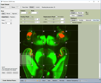
Figure 1: Stitching of individual tiles using the Tile Sorter; shown are 35 individual z-stacks covering a dual labelled mouse brain (CCKBAC/DsRed::GAD67gfp/+) at embryonic day 16.5; GA-BAergic neurons are marked by GFP (green) due to expression of GAD67; CCK expression is marked by DsRed (red). Tile 4 is selected and highlighted by a white box. For aligning, the Tile sorter provides different methods including Grid, Manual and Alignments using advanced algorithms. In this case, the z-stack tiles were sorted using the Grid mode; Pixel overlap was set to 10% as during imaging. Several planes were checked to assure correct alignment. Due to stability during image acquisition no further adjustments as algorith-ms were used. Scale options and transparency settings (see selected Tile 4) help to assure correct alignment.

This study benefitted from arivis  Vision4D as the only software with the ability to easily stitch, visualize and share large data sets. Therefore, only the combination of Light-Sheet Microcopy and arivis Vision4D allowed the researchers to obtain 3D information of the entire brain and share this via movies in a very demonstrative way. Based on this, a classical tangential migratory route of CCK interneurons while populating the cerebral cortex was ob-served. Overall, this study identified several characteristics of prenatal CCK interneurons and integrates important information for unravelling the diversity of interneuron functionality
ABOUT US

ZEISS arivis Scientific Image Analysis

Carl Zeiss Microscopy Software Center Rostock GmbH (formerly arivis AG) is a Germany-based software company specializing in image analysis software solutions for the life sciences industry. Our in-house developed ImageCore Technology lies at the base of our products and enables scientists to work with large datasets using their desktop workstation. ZEISS arivis Pro (formerly Vision4D)  allows users to manipulate and analyze terabyte-sized images with features such as color mapping, rendering, and quantification. For larger scale data analysis, ZEISS arivis Hub (formerly VisionHub) provides a server-based image analysis framework accessible through a standard web browser. The company also offers the arivis Pro VR toolkit (formerly VisionVR), the world's first virtual reality system for microscopy image data, allowing scientists to fully immerse themselves in their samples.

Source Dataset Specifications

Microscope: Lightsheet Z.1 microscope (Zeiss), ×5 (EC Plan Neofluar 5×/0.16) detection objective, ×5/0.1 illumination optics, PCO edge sCMOS camera; Imaging settings: ×0.7 zoom, laser power of 20v%, exposure times of 200 ms, tile scan overlap 10%; Data size: 100-300 GB; Picture size: up to 48 tiles, each 1200 px x 1200 px; Voxel size: 1,3 µm x 1,3 µm x 3,5 µm; z-depth: up to 1500 planes

arivis Software Package

arivis Vision4D Base Package (now ZEISS arivis Pro)