Authors: Bechara Saykali, Navrita Mathiah, Wallis Nahaboo, Marie-Lucie Racu, Latifa Hammou, Matthieu Defrance, Isabelle Migeotte
Researchers at ULB are using ZEISS arivis Pro (formerly Vision4D) to study the very early stages of a mouse embryo development.
This is a research area that can generate large image files as we try to capture bigger fields of view and higher spatial and temporal resolution. Isabelle Migeotte’s group used two-photon microscopy and live imaging to follow and understand the gastrulation process in the mouse embryo. In particular, they focus on the differentiation and cell migration process of cells in the mesoderm layer.
ZEISS arivis Pro (formerly Vision4D) software allowed the researchers to track cell movement and cell morphology over the time series. The paper published by Saykali et al. reports different phenotypes for embryonic and extra-embryonic mesoderm populations depending on the cell fate and 3D environment.
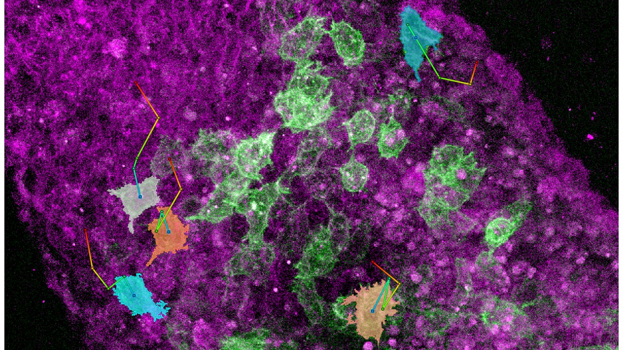
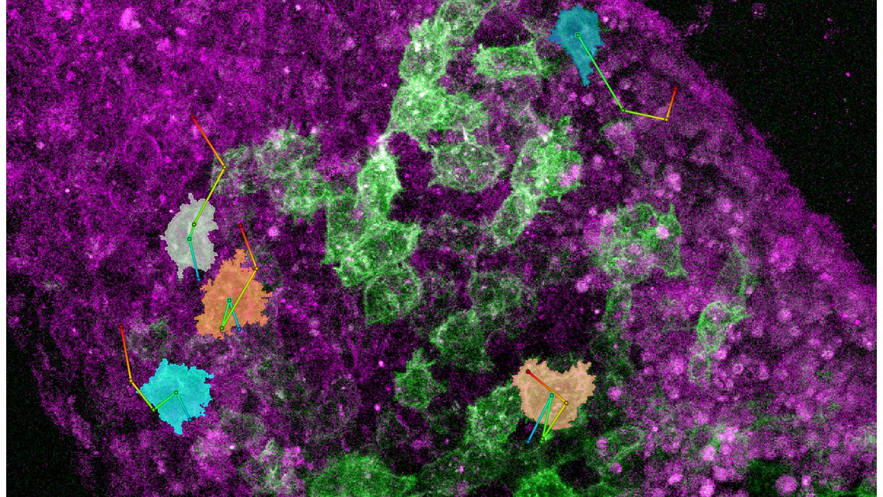
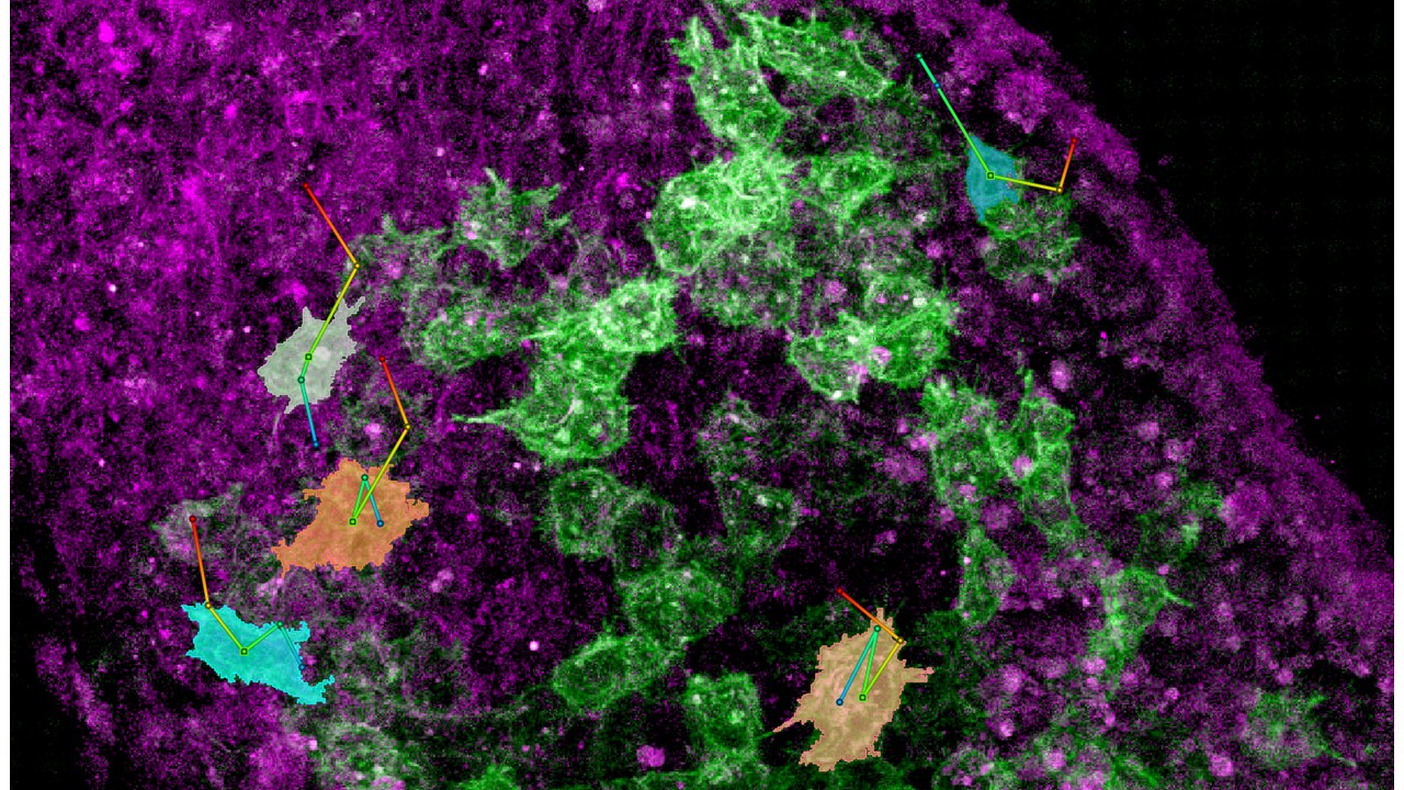
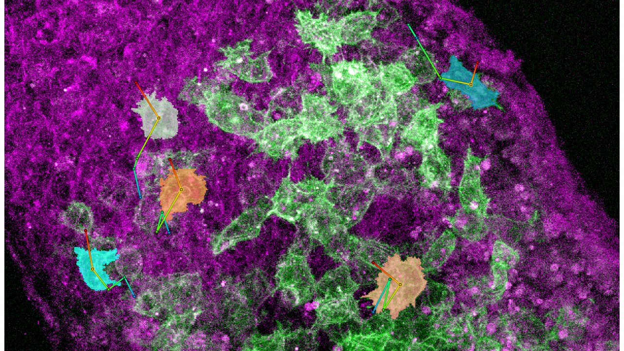
Embryo drifting movement was first corrected by manually segmenting the contour of the embryo, tracking the whole embryo, and using ZEISS arivis Pro (formerly Vision4D) drift correction tools. Due to the complexity of the data, cells of interest were manually segmented and tracked in ZEISS arivis Pro (formerly Vision4D). The researchers quantified cell parameters such as net displacement, track length, speed, angle between two cells, and length of cell filopodia using a combination of ZEISS arivis Pro (formerly Vision4D) and Python scripting (ZEISS arivis Pro - formerly Vision4D allows the user to run and edit Python scripts).
Image Courtesy: Saykali et al. Distinct mesoderm migration phenotypes in extra-embryonic and embryonic regions of the early mouse embryo.
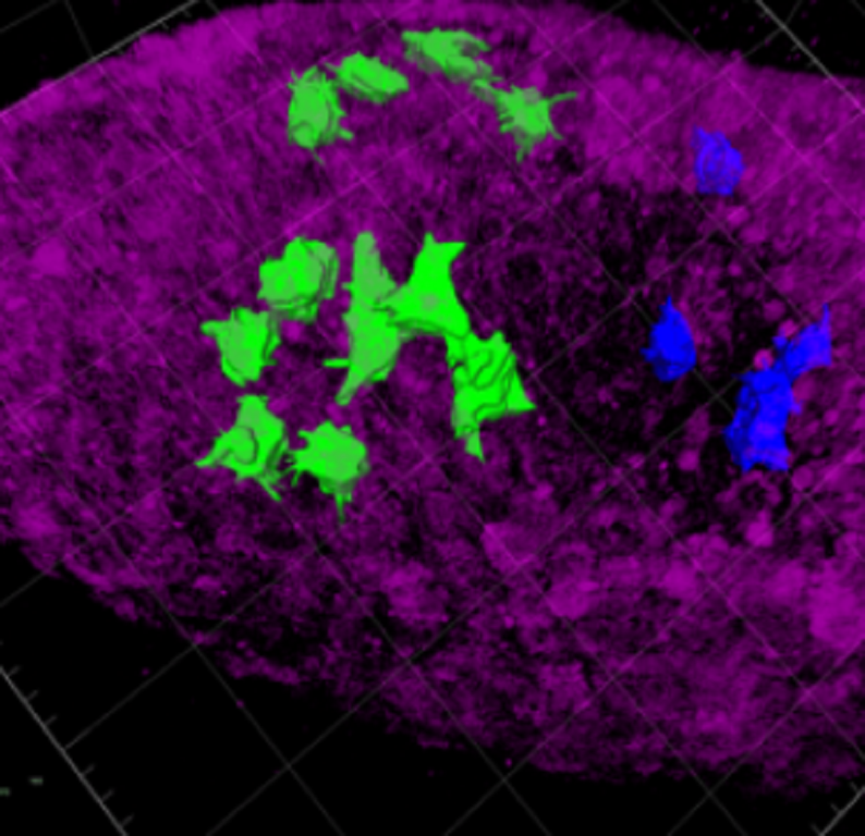
Morphologically, extra-embryonic cells are larger, longer and develop small protrusions (blue colored cells) versus the embryonic cells (green) which are smaller in volume with more protrusions.
On average embryonic cells were approximately 2.000 µm3 in volume with a mean filopodia length of 8 µm. Extra-embryonic cells were double inside (4200 µm3) with shorter filopodia (6 µm).
Image Courtesy: Saykali et al. Distinct mesoderm migration phenotypes in extra-embryonic and embryonic regions of the early mouse embryo.
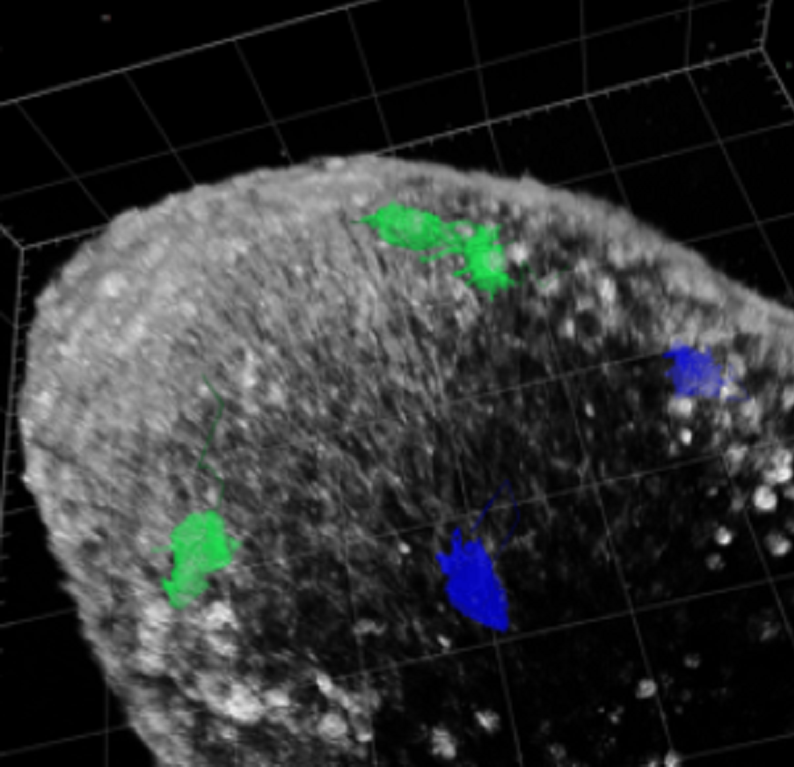
In terms of migration patterns, embryonic cells tend to have a more directional trajectory (green) versus a zigzag movement shown by extra-embryonic cells (blue).
Researchers report that embryonic mesoderm cells moved at a mean speed of 0.65 um/min and travel approximately a distance of 90 um, with a track straightness value of 0.48 (a value of 1 representing a linear path).
Extra-embryonic cells migrated slightly slower at an average speed of 0.45 um/min, travelled shorter distances of around 70 um with a track straightness of 0.3.
Image Courtesy: Saykali et al. Distinct mesoderm migration phenotypes in extra-embryonic and embryonic regions of the early mouse embryo.
3D tracking of cells can be challenging due to:
This tutorial gives you an introduction on how to use ZEISS arivis Pro (formerly Vision4D) to track moving objects in a microscopic time series image.
Click below to see a detailed workflow on how to do time-lapse analysis.
If your sample is drifting, it is best to correct for the drift first.
If you have a poor signal-to-noise ratio, use some preprocessing tools for better cell detection.
Segment operators that tend to work very well are Blob Finder and Watershed.
Use a Segment Filter to exclude any false positive cells or select only certain cells of interest.
Use the Tracking operator to track cells. Linear Regression and Brownian algorithms (depending on whether the movement is directional or not respectively) tend to provide good results.
Research Paper: Bechara Saykali, Navrita Mathiah, Wallis Nahaboo, Marie-Lucie Racu, Latifa Hammou, Matthieu Defrance, Isabelle Migeotte. Distinct mesoderm migration phenotypes in extra-embryonic and embryonic regions of the early mouse embryo. eLife 2019;8:e42434.
DOI: 10.7554/eLife.42434