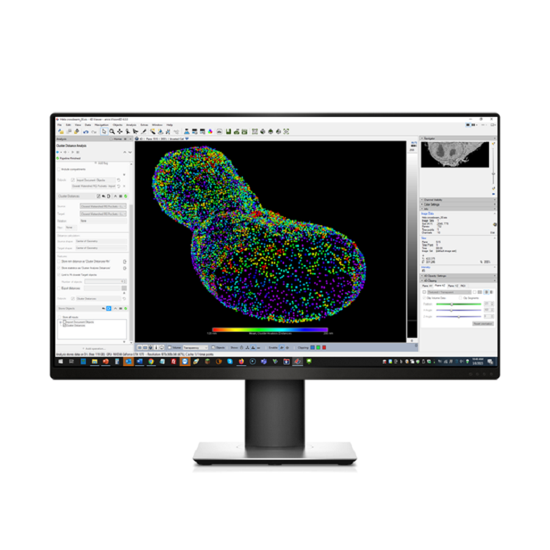
ZEISS arivis Pro is your modular image analysis software when working with multi-channel 2D, 3D, and 4D images of virtually unlimited size. With ZEISS arivis Pro (formerly Vision4D) it is easy to create automated end-to-end image analysis pipelines with just a few clicks. Use predefined pipelines for common use cases or create your own pipelines using the flexible click-and-play interface.
Highly scalable computing, not dependent on local system resources
Processing and quantifying any kind of multidimensional image data
Combining different operators for complex analysis
Denoising, AI-driven segmentation, filtering, thresholding and more
ZEISS arivis Pro 4.2, empowers researchers with unprecedented flexibility to tailor microscopy image analysis to their unique needs.
This major release introduces:
ZEISS arivis Pro 4.2 is a universal solution optimized for any imaging workflow. This release underscores the ZEISS commitment to delivering cutting-edge, user-centric solutions that accelerate your research.
Reconstruction of Segmented Mouse Muscle Ultrastructure. This detailed 3D volume rendering showcases the intricate organization of the key cellular components within mouse muscle tissue.
Sample courtesy of Prof. Guorui Huang, Ruijin Hospital, Shanghai Jia Tong University School of Medicine.
The ZEISS arivis Pro analysis strategy and iterative approach allow processing and segmentation of a small field of view, a 3D/4D subset, or the entire dataset. Analysis results can be viewed in synchronized split windows in 2D and 3D views simultaneously, which is particularly useful for densely packed structures and tracking experiments. The software's integrated Machine Learning/Deep Learning functionality (available as local training option or in the ZEISS arivis Cloud) allows for setting up an image segmentation and analysis pipeline with ease and without extensive knowledge of AI analysis methods.
Analyze complex and challenging samples with a few clicks. No matter the source and format of the image, the arivis scientific image analysis platform is designed to easily process and analyze it. Our software tools are highly integrated and can benefit customers in academia, across industries and various applications, including biotech, pharma, life science and material science.
AI-assisted 3D Density Analysis of a HeLa cell, imaged with ZEISS FIB-SEM Technology
Original datasets imaged with a ZEISS FIB-SEM instrument and kindly provided by Anna Steyer and Yannick Schwab, EMBL Heidelberg, Germany.
ZEISS arivis Cloud was implemented to train neural networks that identify large organelles: mitochondria and the nucleus. Using the arivis AI toolkit on ZEISS arivis Cloud (formerly APEER), we manually annotated a subset of the instances of these cellular features and trained neural-network models that successfully predicted the remainder of the instances of cellular features within the FIB-SEM image volume. These AI-trained models were first used to infer mitochondria and the nucleus in ZEISS arivis Pro (formerly Vison4D). We then built automated analysis pipelines to filter and improve the initial inferences into usable 3D segments.
Overall, this work highlights how the deep learning approach, when combined with the powerful 3D tools of ZEISS arivis Pro, enables 3D segmentation and measurements within FIB-SEM image sets.
Choose the ideal pipelines and modules for your research
Get a new perspective and experience your sample in a whole new dimention with the emersive virtual reality environment, using the arivis Pro VR toolkit.
The arivis Pro Exchange Objects Module allows for easy interface and import of your specific workflows from a third-party or open source, such as FiJI, ImageJ or Ilastik. Import your workflow with a single step and visualize or further analyze your 3D objects with ZEISS arivis Pro. Create high-quality renderings to publish your results. The 2D/3D split views allow interactive exploration of the results or interactive gating in scatter plots or timeline graphs.
The arivis Pro Batch Analysis Module can be used for the unattended execution of a pre-configured analysis pipeline on a series of images or regions of interest. The pipeline is easily adapted and can be applied to multiple datasets.
Automatic Neuron Tracing includes two established scientific methods to choose from. Neuroscientists use this module to generate quick results even with large microscopy imaging data.
The state-of-the-art, integrated algorithms can save you precious time and effort when visualizing and tracing neuroscience datasets. The module offers compatibility and flexibility for a wide range of sample data and imaging modalities.
No matter if your research centers on confocal or light-sheet microscopy, single neurons or cleared tissue applications, Automatic Neuron Tracing includes a wide variety of the most relevant trace measurements. The easy-to-use, high-performance semi-automatic trace editing tool enables easy correction of trace paths even for difficult samples.
arivis Storyboard assists you with export of impressive cinema-quality animations of your results: Let your data speak for itself!
Automated 3D neuron tracing of neuronal tissue (mouse brain) with Vision4D 4.0 and ZEISS LSM 980 confocal microscope. Original 3D tissue volume dataset (45GB CZI, confocal fluorescence microscopy, single channel) kindly provided by Dr. Steffen Burgold, ZEISS RMS Customer Center Oberkochen, Germany.
Leading institutes and research organizations trust ZEISS arivis for scientific image analysis.