Adjust the slider to view the 'before and after' comparison.
Solution Description
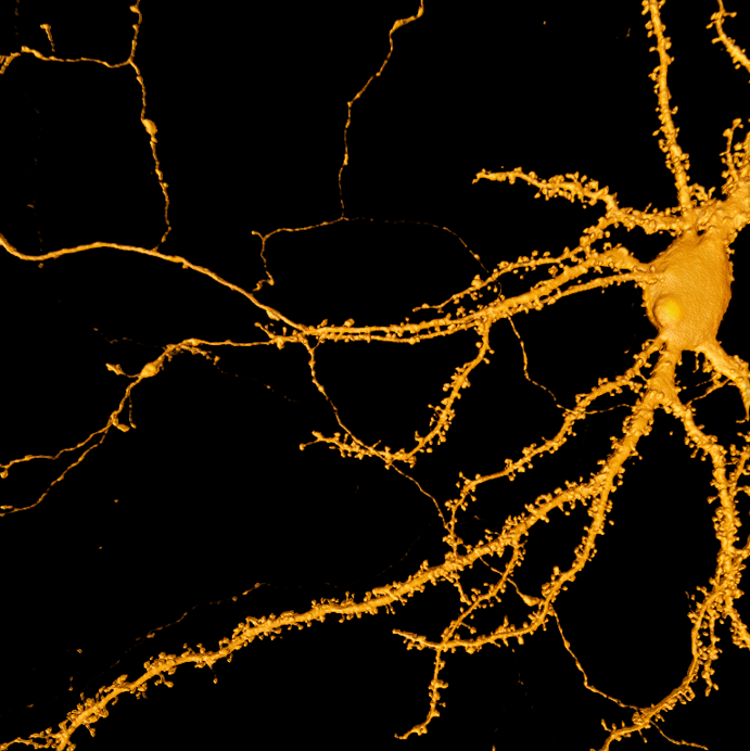
Microscopy and deep learning are valuable tools in Parkinson's research, allowing researchers to study neural circuits and understand the cellular mechanisms that regulate synapse formation and composition.
In this application, a deep learning-based semantic segmentation model was trained using arivis AI to separate dendritic spines and neuronal projections using 3D z-stack images captured from a Zeiss Celldiscoverer 7 microscope. An iterative process involving data-centric model training was employed to refine the model before integrating it into an image analysis pipeline utilizing the 3D toolkit in ZEN.
The successful segmentation of dendritic spines using the trained model demonstrates the effectiveness of deep learning in complex image analysis and its potential to contribute to future neurological disease research.
Keywords:
Deep learning, arivis Cloud, arivis AI, Celldiscoverer, LSM 900, Airyscan, spines, dendrites, neurons, ZEN
Image Source:
Sample courtesy of R. Thomas and D. L. Benson, Icahn School of Medicine, Mount Sinai, New York, USA

.png)
