Adjust the slider to view the 'before and after' comparison.
Solution Description
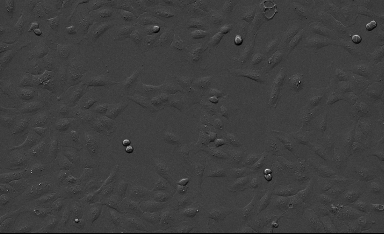
Counting cells is one of the most important and common applications in biology. At the same time, this is a highly challenging task if the cells are not fluorescently labelled or stained. Therefore, classical image processing methods can often not be used for brightfield images.
This solution is based on a pre-trained deep-learning network that detects the cells in one or more bright field microscopy images. Time series images as well as multi-well images are supported. The output is a heatmap that shows the number of cells per image in each well and time point (if applicable). The respective deep-learning network is trained on datasets from multiple microscopes with different resolutions/magnifications.
Figure 1: Number of cells are plotted as a heat map, indicating the number of cells per image across a 96-well plate (red: high, blue: low).
Keywords:
Deep learning, arivis Cloud, cells, counting,brightfield, phase contrast

.png)
.png?width=624&height=400&name=63861162f78b032eaf6ed4b9_Cells-plot%20(1).png)
