
In the joint series "From Image to Results", explore various case studies explaining how to reach results from your demanding samples and acquire images in an efficient way. For each case study, arivis together with ZEISS Microscopy highlighted different samples, imaging systems, and research questions. In this blog article we will present the highlights of 2021 that will inspire your own research in scientific imaging.
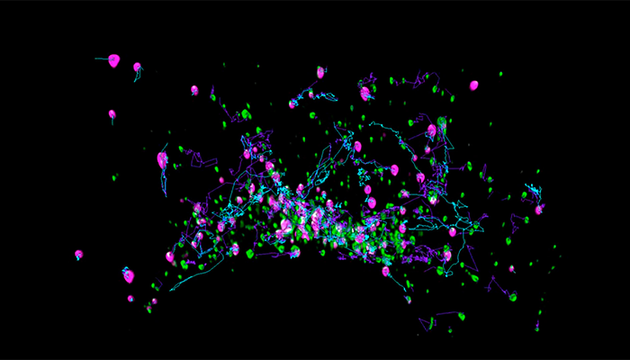
Sample: Cos7 cells transiently transfected with mEmerald-Rab5a and Golgi7-tdTomato, Task: Track both types of vesicles, volume & distances over time, colocalization, Results: Graphic visualization of tracks and cells in a movie and resulting data as time line along track, scatter plots, System: ZEISS Lattice Lightsheet 7, Software: arivis Vision4D.
Vesicle Trafficking in cos7 Cells:
We start the series with an analysis of vesicle trafficking in Cos7 cells. The goal is to generate a high-resolution longitudinal study of vesicle trafficking within a single cell to explore some basic observations concerning vesicle transport. We then showcase image analysis tools to segment vesicles and generate vesicle tracks to derive meaningful data to confirm our observations.
Vesicle Trafficking with Vision4D
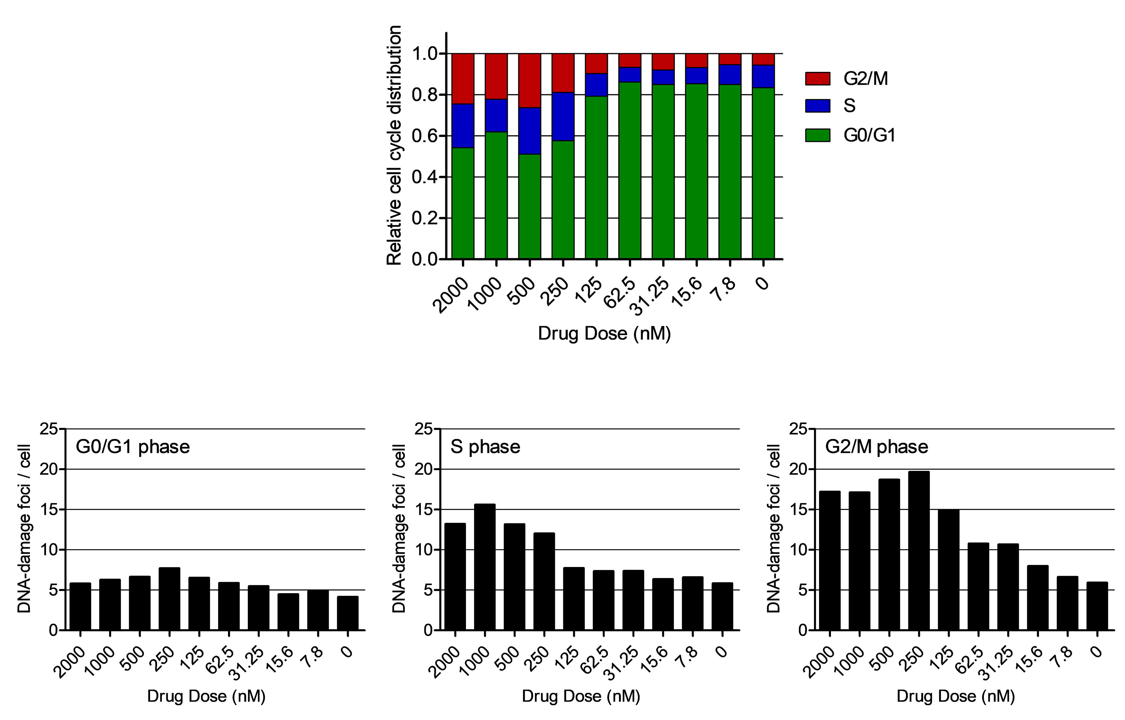
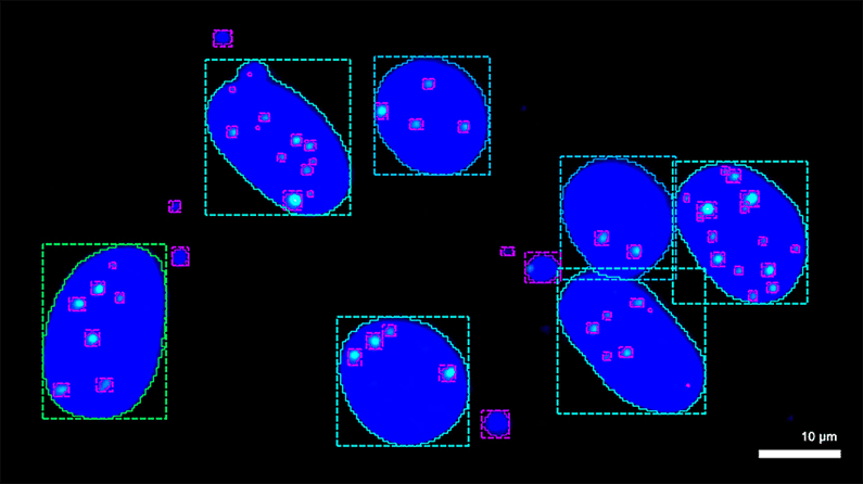 Sample: Clear-cell renal carcinoma cell line SLR22, Task: DNA-damage analysis, Results: Cell-cycle specific DNA-damage elucidates drug mechanism, System: ZEISS Axio Observer, Software: arivis Vision4D, arivis VisionHub.
High Content Imaging for Genotoxicity:
In this second episode, we set up an application example to perform DNA-damage analysis by employing ZEISS Axio Observer and the arivis Vision4D image analysis software. We highlight the general analysis as performed with one drug. The aim is to show how to automate imaging and analysis procedures, to the point that the whole process can be scaled easily to perform high-content screening (HCS) or be adapted to other complex experimental settings.
High Content Imaging WITH VISION4D
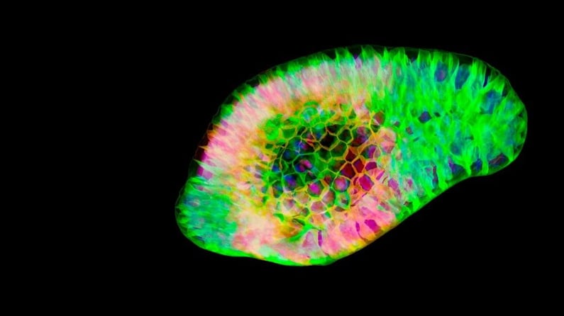 Sample: Intestinal organoids, Task: Study the role of Wnt signaling in organoid formation, Results: Analysis of an organoid experiment, System: ZEISS Celldiscoverer 7, Software: arivis Vision4D, arivis VisionHub.
Organoid Analysis:
In this third episode, we showcase a simple imaging experiment performed on intestinal organoids treated with and without a Wnt-inhibiting drug, with the experimental goal to study the role of Wnt signaling in organoid formation. Once a pipeline has been created and optimized in arivis Vision4D by testing on a sample set of images, it is then possible to scale up the analysis by use of the server-based VisionHub platform. As arivis VisionHub is accessed over the web, users can easily upload pipelines to their datasets, run batch analysis and then have the results readily available within the web-based user interface.
ORGANOID ANALYSIS WITH VISION4D
We hope that you find these case studies interesting and inspiring in your own research. If you'd like to talk about any of these studies with an arivis expert we invite you to request a personal remote demo. Of course you can also register for your personal free trial version of Vision4D and see for yourself what our scientific imaging platform can do with your scientific datasets - we support a vast selection of file formats and systems.
We will continue with this exciting series in 2022 - see you next year!