
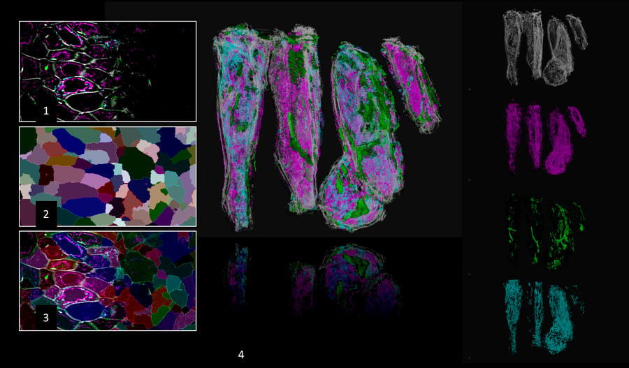
With arivis you can now integrate all tasks of image visualization and analysis of multidimensional microscopy imaging data of any size in a single software package.
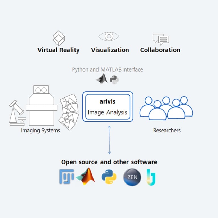
The Open Source community offers many special functionalities and regularly adds new ones to the research community. On the other hand many users prefer a commercial, robust and fully supported image analysis platform with a unified UI that covers all aspects of image processing, analysis and visualization of results via desktop workstation, cloud computing or Virtual Reality. Therefore an easy transition between open source/third party packages and our arivis Scientific Imaging Platform is essential.
With the ZEISS arivis Pro Exchange Objects with Open Source module we offer an integrated solution to imaging facilities and PIs to connect with open source workflows as well as with any other software which is capable of exporting/importing labelled images. Easily interface with your specific workflows whenever you are using segmentation functions with open source packages such as Fiji, ImageJ, Ilastik, Python or with commercial solutions such as MATLAB or ZEN/APEER software by ZEISS Microscopy.
No programming knowledge needed - ZEISS arivis Pro adapts to your requirements and workflows!
Perform segmentation with your preferred scientific open source or commercial package

The arivis Scientific Image Analysis Platform is a flexible computing universe that scales, parallelizes, and streamlines all imaging workflows. With arivis' easy sharing functions your organization can enjoy data proficiency and efficiency at new levels. arivis includes integrated toolsets that handle everything, from file storage format to project and user-specific computations, to reporting. The platform's hubs connect datasets and manage central imaging databases and raw data exposure, while also enabling Machine Learning and AI routines.
Extract results from your scientific images with ZEISS arivis Pro. Make the most of hardware power adjustments and smooth interactivity on microscopy datasets of virtually unlimited size. The software unites multiple tools for visualization and advanced analysis into one easy-to-learn user environment for high productivity. Extend your possibilities with the Python coding feature and a multitude of supported libraries and APIs. Optional Virtual Reality interface.
