Nanoparticles research plays a very important role in numerous industrial applications such as pharmaceuticals, biomedical applications, coatings, inks and pigments, energy materials and filtration. To engineer nanoparticles with unique properties, improve synthesis methods and innovate new products, the chemistry, size and shape of individual nanoparticles must be characterized. Even though there are bulk analytical techniques (such as sieving or laser scattering) to determine particle size distribution (though these methods may be limited by particle size and/or composition), auto-mated analysis of individual nano particles in agglomerates still remains a challenge.
As traditional image segmentation algorithms fail to identify the boundaries between individual nanoparticles, this type of investigation is still performed by a human operator and often leads to inaccurate and inconsistent results.
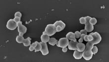
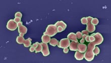
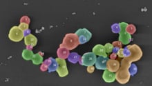
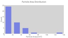
Figure 3 Workflow of nanoparticles size distribution analysis. (A) Original Scanning Electron Microscope (SEM) image of nanoparticles acquired at 2 kV using the Inlens detector. (B) Image of segmented image using Intellesis showing background (blue), boundaries between particles (green) and nanoparticles (red). (C) Image of separated individual nanoparticles using machine learning and further analysis. (D) Particle area distribution of individually segmented nanoparticles
Figure 3 shows an example of an end-to-end automated workflow used to separate individual nanoparticles in agglomerates and to determine their particle area distribution. Nanoparticles from the sparks of ferrocerium collected on a silicon substrate were acquired using Secondary Electron detector, on a ZEISS GeminiSEM 500.[5,6] Machine learning segmentation in ZEISS ZEN Intellesis was successfully used to identify three different classes: nanoparticles, boundary between nanoparticles and background. To further separate individual nanoparticles and determine the size distribution, an open-source python package (https://scikit-image.org) was created in arivis Cloud, the cloud-based digital microscopy platform for ZEISS (see below), for a seamless and personalized analysis. Using an automated workflow that combines advanced microscopy, machine learning image segmentation and analysis, characterization of individual nanoparticles in agglomerates was performed. This type of investigation helps researchers to better understand the relationship between material properties to further advance industrial research.
