Integrating subcellular information into the context of whole tissues is important for understanding cellular networks and their contribution to normal and diseased conditions. Challenged by the size and opacity of tissues, only recent advancement in imaging techniques paved the way to study intact organs at a subcellular resolution. However, using techniques like Light-Sheet Microscopy is accompanied by the generation of large image data files, which complicates post-imaging data handling and processing drastically.
The acute brain injury research group of Dr. Ali Ertürk at the Institute of Stroke and Dementia Research (ISD) in Munich, is leading in breaking the limitations of volumetric imaging by using Light-Sheet Microscopy combined with innovative tissue clearing methods. Tackling the challenges of large imaging data sets, the lab benefitted from the unique feature of ZEISS arivis Pro (formerly Vision4D) as an imaging data platform for easy handling and processing of data sets unlimited in size.
Based on the arivis ImageCore Technology, ZEISS arivis PRo (formerly Vision4D) is unreached in its performance of handling large imaging data sets even on standard hardware. This product combines a variety of data pre-processing features and interactive visualization tools with the ability to handle large data sets, giving the user the possibility to standardize complete workflows.

Dr. Ali Ertürk
Institute for Stroke and Dementia Research (ISD), Klinikum der Universität München, Acute Brain Injury Research Group
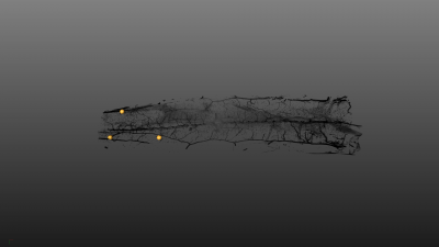
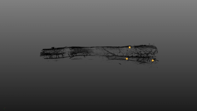
Figure 1: Fusion of separate mouse spinal cord data sets. arivis Scope fusion allows the fast combination of large data sets with different orientations (upper row) into one data set (see above).
In this study, Dr. Ali Ertürk and his team analyzed the mouse spinal cord using Light-Sheet Microscopy (LaVision Biotec, Ultramicroscope II) and uDISCO tissue clearing (https://www.ncbi.nlm.nih.gov/pubmed/27548807). Due to sample size, image acquisition was done via mosaic tile scanning resulting in three individual stacks with different z-orientations. This imaging procedure necessitated a demanding post-imaging processing in order to fuse the individual stacks into one complete data set of the spinal cord. This process was further complicated by the size of one data set (300 GB per volume). Only the interactive Scope fusion tool of ZEISS arivis Pro (formerly Vision4D) was able to fuse these large data sets to one resulting image 1,2 TB and open the possibility to study the spinal cord in its native structure.
Carl Zeiss Microscopy Software Center Rostock GmbH (formerly arivis AG) is a Germany-based software company specializing in image analysis software solutions for the life sciences industry. Our in-house developed ImageCore Technology lies at the base of our products and enables scientists to work with large datasets using their desktop workstation. ZEISS arivis Pro (formerly Vision4D) allows users to manipulate and analyze terabyte-sized images with features such as color mapping, rendering, and quantification. For larger scale data analysis, ZEISS arivis Hub (formerly VisionHub) provides a server-based image analysis framework accessible through a standard web browser. The company also offers the arivis Pro VR toolkit (formerly VisionVR), the world's first virtual reality system for microscopy image data, allowing scientists to fully immerse themselves in their samples.