arivis Scientific Image Analysis

Advanced 4D Tracking of Meiotic Spindle Dynamic

Request a Trial
CASE STUDY

Live-cell Imaging of Meiotic Events in C. elegans

With its expertise on correlative light and electron microscopy (CLEM), the laboratory of Prof. Dr. Thomas Müller-Reichert at the TU Dresden is committed to study the details of meiotic cell divisions and the function of the spindle apparatus during this process using the model organism C. elegans. Whereas mitotic spindle dynamics have been studied in detail within C. elegans, the dynamics during male meiosis remain mainly elusive. Using the live animal, Gunar Fabig, PhD student at the Müller-
Reichert lab, conducted for the first time a quantitative analysis of spindle dynamics during male meiosis I and II in 4D. Combining the advantages of C. elegans (transparency, easy mounting, the possibility to fluorescently tag proteins, accessibility of the gonad) with spinning disc microscopy (fast image acquisition), Gunar was able to obtain 3D stacks of the full male gonad every 30 s for 45 to 60 min.

2000px-Logo_TU_Dresden-300dpi-min

Gunar Fabig, Müller-Reichert lab, Experimental Center, Medical Faculty Carl Gustav Carus, Technische Universität Dresden (TUD), Germany.

Download print version

Tackling Challenges in Image Analysis

Analyzing the spindle pole dynamics of wild-type C. elegans in these data sets was quantitatively approached by measuring the spindle pole-to-pole distance over time. By doing so, the researchers were confronted with two major challenges. First, due to 4D imaging of live animals, every spindle was randomly located and orientated within the data set. Second, although mechanically immobilized, a slight movement of the roundworm could not be prevented. These conditions necessitated a demanding image analysis including 4D segmentation, 4D tracking and the possibility to correct tracking failures as a consequence of sample movements.Combing his imaging approach with ZEISS arivis Pro (formerly Vision4D), allowed Gunar to tackle the challenges, extract the desired information in an efficient manner and develop a standardized analysis pipeline. He specifically benefitted from the possibility to analyze randomly oriented spindles by calculating the center of mass of the spindle poles and to edit wrong tracks very easily with the track editor.
Using this interactive tool, individual tracks could be easily identified, selected and edited, which includes merging or splitting of tracks by a simple drag and drop function.

Analysis Pipeline Using arivis

Based on these advantages, Gunar Fabig created an analysis pipeline within ZEISS arivis Pro (Formerly Vision4D) to standardize the analysis of spindle elongation:

  • After import, a denoising filter was applied.
  • Individual meiotic events within the gonad were analyzed separately by creating 3D Regions of interest to export single spindles.
  • Within these ROIs, spindle poles marked by GFP were defined in 3D using an intensity threshold segmentation.
  • To exclude small, false positive segments, a filtering step based on volume size was applied.
  • The segmented spindles were tracked over time by using the arivis tracking module including the track editor.
  • The analysis report was exported as Excel file including the center of mass coordinates of each segment over time.

Towards a Big Picture of the Meiotic Spindle

Track editor_neu-min

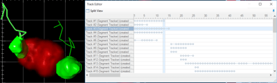
Figure 1: 4D segmentation and 4D tracking of spindle dynamics; (left) 3D representation of male meiosis I in living C. elegans; spindle poles are marked by GFP fused to γ-tubulin, chromosomes are marked by mCherry fused to histone H2B. Segmented spindle poles are shown in green, chromosome are shown in red using the 4D Viewer of ZEISS arivis Pro (formerly Vision4D). Tracks of segmented spindle poles over time are depicted as lines. The left track is selected and can be easily identified using the track editor (right), highlighted in blue. Within the track editor, individual tracks are shown on the left, time frames on the top. Every segment is visualized as a small circle. A series of connected segments that form a track is visualized by a horizontal line connecting the segment circles. Merging or splitting of tracks can be easily done by clicking on segments and dragging them to their desired position.

Based on this fast, quantitative analysis of spindle dynamics in wild-type C. elegans using ZEISS arivis Pro (formelry Vision4D), the Müller-Reichert lab is now able to further complete the big picture of the meiotic spindle and address outstanding questions. Altogether, this analysis will shed light onto the molecular mechanism, which allows the spindle apparatus to accurately segregate paired and unpaired chromosomes.

Download print version

Source Dataset Specifications

Microscope: Olympus IX83, invers; Andor IQ 3.2; equipped with Yokowaga CSU-X1, Andor iXON Ultra 897 EMCCD detection camera and Olympus U Plan S Apo 60x / 1.2 W objective; Data size: 1 GB to 3 GB; Time series: stack acquisition every 30 s for 45 to 60 min; Picture size: 512 x 512 px; Voxel size: 0.222 µm x 0.222 µm x 0.33 µm; z-depth: ~60 planes, 19,8 µm;

Software Package

ZEISS arivis Pro (formerly Vision4D)