TILING & STITCHING

Ask for a Trial

Stitching & Alignment

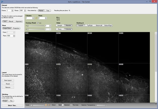
Various applications require a process named stitching and alignment as a prerequisite step for the visualization of 3D or 4D volumetric files. Here, every plane of the resulting image is combined of a number of tiles acquired separately.
arivis Vision4D is the unique tool for effective stitching and alignment of hundreds to thousands of multiple single files (tiles), which may create volumes of several hundred gigabyte or even terabyte in total file size.
The arivis Tile Sorter allows for a complete interactive adjustment for stitching.

Talk with an Expert

arivis Vision4D-Tile-Sorter screenshot

Features

  • Arrange single and isolated image tiles to a large stitched volume
  • Plane shift for tile stacks
  • Supports source data with tiles organized as scopes
  • Support for manually rotated tiles
  • Manual as well as automatic stitching and blending
  • Blending between overlapping regions
  • Methods for aligning
  • Grid: Manually define order & overlap based on equally distributed grid
  • Manual: Drag and drop single tiles by hand
  • Alignment: Advanced algorithms will find and align matching tiles
  • Optional maximum Intensity Projection (MIP) mode to improve results
  • Import, review & stitching of TrakEM2 projects
  • Powerful Alignment algorithms and optimizers
  • Phase Correlation Method (PCM)
  • Normalized Cross Correlation Method (FFT-NCC)
  • Template matching alignment
  • Energy optimizer
  • SVD optimizer

Courtesy of M. Paoli, Galizia Lab, University of Konstanz, Germany, Image acquisition with ZEISS LSM with Airyscan
Image Details: The brain, thoracic and abdominal ganglia of the cockroach are joined together by bilateral connective bundles of ascending and descending interneurons forming the ventral nerve cord. In this preparation, left and right connectives were individually labelled (Alexa 488: green, Alexa 647: magenta) posteriorly to the suboesophageal ganglion to observe the extension of their innervation within the different neurophils, and throughout the ipsi- and contralateral parts of the brain (DNA labelled with DAPI: cyan). Imaging was performed using Tiling and Stitching to capture the complete volume (3×2.3× 0.26 mm). 3D animation of the complete dataset was done with arivis Vision4D, ideal for rendering and analyzing large datasets. The 4D viewer in arivis Vision4D can be configured to adjust the appearance of individual channels independently to highlight specific features.

The arivis Imaging Platform

The arivis Imaging Platform is a flexible computing universe for Imaging Science that scales, parallelizes, integrates and connects all imaging workflows, sparking organization-wide image data proficiency and efficiency at all levels. The integrated toolsets take care of everything from the file storage format to user and project-specific computations to reporting. The computational and management hubs that comprise the platform connect your datasets and take care of your central imaging databases and can expose data assets - including raw data and specified portions of raw data - to Machine Learning and AI routines.

OUR TEAM IS READY TO HELP

Use the form below to get in contact with our arivis experts. Please describe your query and our team will contact you.