
With Machine Learning and Deep Learning tools available in the ZEISS arivis scientific image analysis eco-system, segmentation of multi-channel images becomes quick and easy.
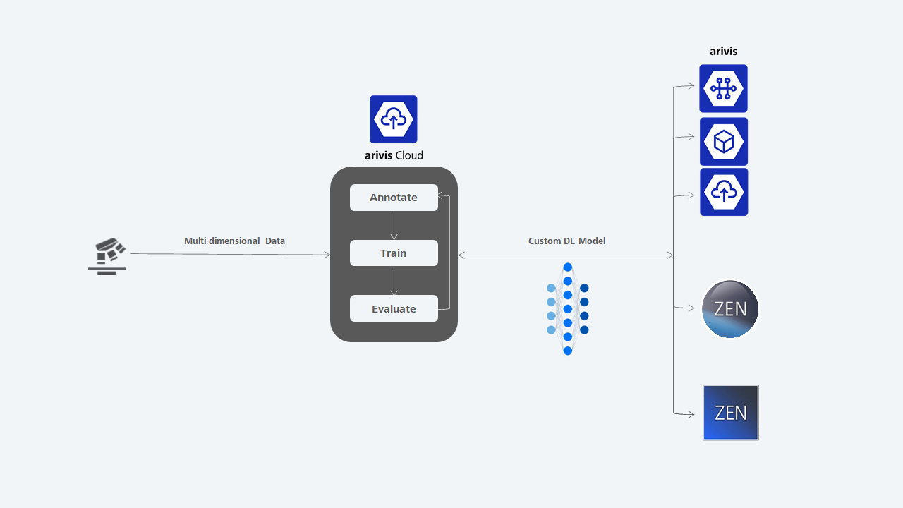
Your scientific expertise is enough to mark and classify structures of interest in your samples. Let The AI toolkit in the cloud train your neural network, then export your AI-trained model. Use the model to set up an end-to-end pipeline with a few clicks.
With arivis machine learning and deep learning tools, you get reliable results in hours instead of weeks or months.
.webp?width=768&height=768&name=AI-ML-DL-graph%20(1).webp)
Are you ready for more AI-driven workflows?
To login to your account, click to go to the login page.

When an imaging specialist designed a conventional algorithm it answers one specific question. A Machine Learning algorithm can be adapted to various questions by training it. The Machine Learning algorithm “learns” patterns and adapts itself.
The researcher need only annotate regions of interest in a small part of the image to train the model. By learning patterns, Machine Learning and Deep Learning tools learn to classify samples, and this information can be then used to identify structures in the entire image and also in other images.
Learn about multiple ways to use your AI-trained model.
The arivis Scientific Image Analysis Platform is a flexible computing universe that scales, parallelizes, and streamlines all imaging workflows. With arivis' easy sharing functions your organization can enjoy data proficiency and efficiency at new levels. arivis includes integrated toolsets that handle everything, from file storage format to project and user-specific computations, to reporting. The platform's hubs connect datasets and manage central imaging databases and raw data exposure, while also enabling Machine Learning and AI routines.
Extract results from your scientific images with ZEISS arivis Pro. Make the most of hardware power adjustments and smooth interactivity on microscopy datasets of virtually unlimited size. The software unites multiple tools for visualization and advanced analysis into one easy-to-learn user environment for high productivity. Extend your possibilities with the Python coding feature and a multitude of supported libraries and APIs. Optional Virtual Reality interface.
