ZEISS arivis Pro is your modular image analysis software when working with multi-channel 2D, 3D, and 4D images of virtually unlimited size. With ZEISS arivis Pro (formerly Vision4D) it is easy to create automated end-to-end image analysis pipelines with just a few clicks. Use predefined pipelines for common use cases or create your own pipelines using the flexible click-and-play interface.
![]() Highly scalable computing, not dependent on local system resources
Highly scalable computing, not dependent on local system resources
![]() Processing and quantifying any kind of multidimensional image data
Processing and quantifying any kind of multidimensional image data
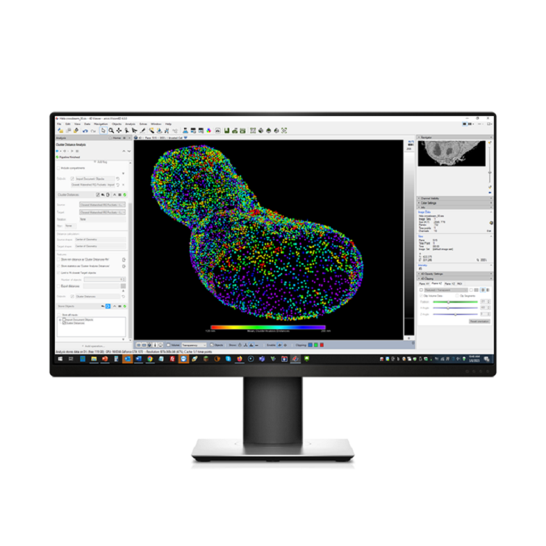
![]() Combining different operators for complex analysis
Combining different operators for complex analysis
![]() Denoising, AI-driven segmentation, filtering, thresholding and more
Denoising, AI-driven segmentation, filtering, thresholding and more

Choose the ideal pipelines and modules for your research
Leading institutes and research organizations trust ZEISS arivis for scientific image analysis.