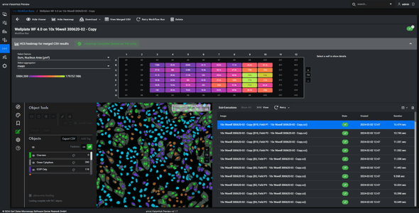
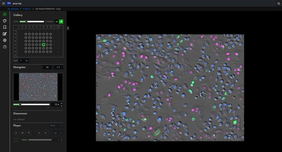
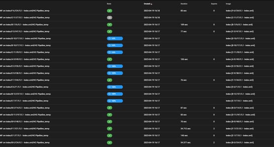
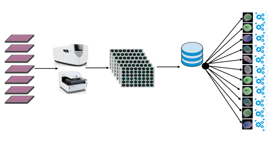
arivis Scientific Image Analysis

High Content Screening and Analysis

Research Process

Scaling Up Innovation

High-Content Screening (HCS) has played a significant role in infectious disease research and drug discovery to date and can be a strong tool in the age of COVID-19 and beyond. With a focus on viral diseases, HCS has been used by laboratories around the world to help with three key interventional strategies: drug re-purposing to fast-track a previously-approved drug to treat new diseases; development of new therapeutics; and vaccine development.

Contact us

High-Content Analysis for Complex Biology

In contrast to the luxury of screening against a critical single target protein, many of the complex diseases that you need to screen drugs against require a broader view of cell or tissue phenotype. This has led to a significant shift towards multi-dimensional High-Content Analysis (HCA) with spheroids, organoids or tumoroids. There are new and emerging important applications involving High-Content Screening that require sophisticated image analysis methods, scalable from 2D to 3D and even 4D.

The arivis Scientifc Imaging Platform allows for highly scalable and automated assay analysis pipelines on the local workstation as well as server-based or cloud-based processing services. Fully integrated workflows for AI methods such as Machine Learning and Deep Learning can save valuable time for recurring and reproducible imaging analysis tasks. Large data sets from past experiments have typically been safely archived and can be re-analyzed with these new methods to generate new discoveries, thanks to the compatibility with multiple vendor-specific file formats.

Fast track approval


WEBINAR

Organoid Image Analysis with the Power of AI to Boost Your Research

In this webinar, you will learn:

  • How to train an AI model to segment your images
  • How to combine your trained model with simple measurements and visualizations with just a few clicks to create a custom application for your repetitive image analysis task
  • How to monitor organoid growth in an automated way in a multi-well format
  • How to analyze organoids in 3D and set up pipelines for complex image analysis
  • How to scale up the analysis to batch process hundreds of 3D organoid images
Talk to an Expert

High-Content Imaging & Analysis

HCS_HCA_ZEISS_arivis_pharma_drug_discovery_infochart


1. Integration Flexible integration of robotics and external hardware via instrument manufacturer APIs.
2. High-content Imaging High-content imaging with manufacturer acquisition software and transfer into arivis.
3. Local HCA & AI Workstation-based imaging assays with powerful ZEISS arivis Pro E2E analysis pipelines for rapid insights.
4. Server HCA & AI Scalable, high-performance analysis, remote access and collaboration with ZEISS arivis Hub.

Computing Infrastructure

ZEISS arivis Hub is a highly scalable and customizable solution that can be adopted to a wide variety of existing data storage and processing infrastructure, from single department and core facility to campus-wide deployment and distributed data centers for multiple locations in pharma industry. However, there is a rising need for out-of-the-box solutions for image storage and processing particularly for small to mid-size businesses such as startups in biotech, CRO, or newly established academia spin-offs that require cost-efficient, modular solutions with minimal administrative hassles.

This is why arivis recommends the HIVE® modular server platform by ACQUIFER™ for rapid and efficient ZEISS arivis Hub deployment. Designed for the pragmatic, biology-focused scientist, the HIVE platform boosts productivity in microscopy facilities and screening labs. HIVE is a high speed centralized data repository that removes the need to constantly move or duplicate data sets in processing and analysis workflows. Its modular design integrates high speed processing, visualization, remote access, project management, flexibility, data security, scalability and ease of use in one unit.

It saves time, space, costs and administrator workload.


Industries Served by the arivis Image Analysis Platfrom

Biotech

3D cell and tissue culture technologies are used more frequently to work with a living organism. 3D cell cultures are replacing the use of expensive animal models. To view and analyse these complex structures arivis developed software tools to help scientists to act faster and get more accurate results.

Pharma

To discover, develop and produce drugs is a time-critical yet complex process, on which  lies the success of a whole industry sector. arivis image analysis platfrom is helping to shorten the evaluation of a new drug by delivering professional, scalable software to analyse and view complex datasets.

CRO

Contract Research Organisations (CRO) provide the pharmaceutical, biotechnology and medical device industries their outsourced services. CROs often need to collaborate and share data with their industrly partner. arivis helps researchers to enhance efficiencies in thier collaboration and research. 

CMO

Contract Manufacturing Organizations (CMO) need professional and scalable imaging analysis to provide results in the quality level required for the pharmaceutical industry today. arivis developed compliant image analysis software tools to fulfil this high demand for industry standards of quality control and assurance.

arivis Pro

Extract results from your scientific images with arivis Pro. Make the most of hardware power adjustments and smooth interactivity on microscopy datasets of virtually unlimited size. The software unites multiple tools for visualization and advanced analysis into one easy-to-learn user environment for high productivity. Extend your possibilities with the Python coding feature and a multitude of supported libraries and APIs.

Advanced AI-driven image analysis
Tile stitching
4D tracking & lineage
Data transformation
Segmentation
Image preprocessing & enhancement
Interactive rendering of volumes and time-series
Cinematic high-resolution rendering
Batch application of analysis pipelines
Custom computed features of objects
Explore results overlaid on raw data

arivis Hub

A nucleus of computational activity and collaboration at scale. arivis Hub enables design and execution of large-scale experiments that produce results from images, whether datasets are already stored or are actively being produced. Mass quantities of microscopy data are onboarded and processed by a virtual team of computational workers. Smartly utilize processing cores in servers or workstations with maximum possible speed.

Register and browse image and data
Manage access for users and groups
Import and archive datasets
Scale your experiments
Leverage computational resources onsite and offsite
Scalable pricing model to support bursts of throughput
Remote rendering servers
Increased QA, efficiency and validity
Smart optimization of parallelization of massive image volumes

arivis Cloud

Train Deep Learning models to automate your image analysis in arivis Cloud (formerly APEER). Whether you use standard pipelines for routine tasks or create your own application, you can power your research with AI with just a few clicks. You can train your own model or alternatively, select a pre-defined pipeline and use a previously trained model on your images. The AI-trained models can be used with other ZEISS software products.


Remote computation resources on the cloud
Easy collaboration and sharing of images and workflows with peers
Deep Learning models are made accessible to everyone
No dependency on an image specialist, intuitive tools for creating your workflows with arivis AI toolkit
No coding required
Improve reproducibility, repeatability and throughput
Reliable results faster
Support from our experts and the image analysis community
RESOURCES

Scientific Applications and Assays

Collected image analysis applications and assays for research microscopy.
Discover the power of the arivis Image Analysis Platform with real-world applications!